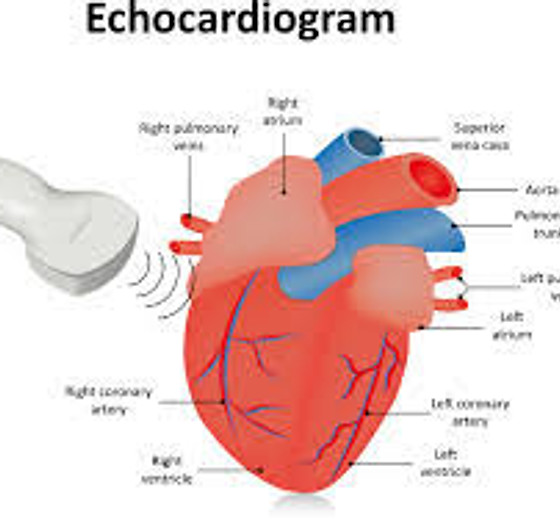
INTRODUCTION OF ECHO
Echocardiograph (ECHO) is a diagnostic procedure of the heart that uses ultrasound waves to take pictures of the heart.
Types of echocardiograph:
Transthoracic echocardiogram.
Transesophageal echocardiogram.
ما هو تخطيط صدى القلب
تخطيط صدى القلب هو فحص للقلب باستخدام تقنية الموجات فوق الصوتية التي تعمل على أخذ صورة للقلب.
انواع تخطيط صدى القلب:
١/ تخطيط صدى القلب عبر الصدر
٢/ تخطيط صدى القلب عبر المريء
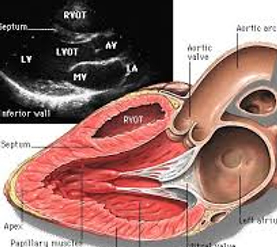
FINDING OF ECHOCARDIOGRAPH
Size and shape of the heart.
Thickness and movement of the heart's wall.
Function of heart's valve.
Find any abnormality in great vessels which inter and leave the heart.
Diagnosis of any abnormality in the heart such as valvular stenosis, valvular regurgitation, congenital heart disease or blood clot inside hearts chambers.
نتائج تخطيط صدى القلب
حجم وشكل القلب
سماكة وحركة جدار القلب
وظيفة صمامات القلب
ايجاد تغيرات غير طبيعية في الأوعية الدموية الرئيسية للقلب
تشخيص أي شذوذ/ خلل في القلب مثل، ضيق في الصمامات، ارتجاع في الصمام، جلطة دموية داخل غرف القلب او عيوب خلقية
HOW DO THE ECHO WORK
The patient lies on a table and a technician places small metal disks (electrodes) on their chest. The disks have wires that hook to an electrocardiograph machine. An electrocardiogram (ECG or EKG) keeps track of the heartbeat during the test. The room better be dark so the technician can see the video monitor properly. Then the technician will put gel on the patient chest to help sound waves pass through the skin. Moreover, the technician may ask to move or hold the patient’s breath briefly to get better pictures.
The probe (transducer) is passed across the patient’s chest. The probe produces sound waves that bounce off the heart and “echo” back to the probe. The sound waves are change into pictures and displayed on a video monitor. The pictures on the video monitor are recorded so the doctor can look at them later.
ماذا يحدث خلال اجراء تصوير صدى القلب
يستلقي المريض على طاولة ويضع اخصائي تقنية القلب أقراص معدنية صغيرة (أقطاب كهربائية) على صدره. تحتوي الأقراص على أسلاك ترتبط بجهاز تخطيط القلب. يتتبع مخطط كهربية القلب (ECG / EKG) نبض القلب أثناء الاختبار. يفضل أن تكون الغرفة مظلمة حتى يتمكن اخصائي تقنية القلب من رؤية شاشة الفيديو بشكل صحيح. ثم يضع اخصائي تقنية القلب مادة هلامية على صدر المريض للمساعدة في تمرير الموجات الصوتية عبر الجلد وأيضا قد يطلب اخصائي تقنية قلب تحريك أو حبس أنفاس المريض لفترة وجيزة للحصول على صور أفضل. يتم تمرير محول الطاقة (transducer) عبر صدر المريض. ينتج محول الطاقة موجات صوتية ترتد عن القلب وتعيد الصدى إلى محول الطاقة. يتم تغيير موجات الصوت إلى صور عن طريق محول الطاقة ثم عرضها على شاشة فيديو ويتم تسجيل الفيديو في الجهاز حتى يتمكن الطبيب من النظر إليها لاحقًا.